 |
 |
 |
Helping You To Achieve ©™

|
![]()
Helping You To Achieve ©™
|
|
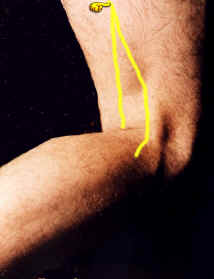
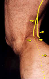
Mark with a surface marking pen the course of the Tibial nerve in the Popliteal fossa and its continuation as the Posterior Tibial Nerve in the Leg on a model in the Anatomical position with a time limit of one minute.
Objective No 159 - Criteria to be demonstrated. 1. Request model to flex the Knee joint to a right angle. 2. Identify and mark the boundaries of the Popliteal fossa. 3. Identify and mark the Medial Malleolus and Medial Tubercle of Calcaneus. 4. Draw a line connecting the Medial Malleolus to the Medial Tubercle of Calcaneus. 5. Mark the mid-point of the line drawn in step 4. 6. Draw a line from a point just above the Apex of the Popliteal fossa, down the midline of the Popliteal fossa. 7. Continue the line on the posterior surface of the leg inclining towards the medial aspect of the heel to join the mid-point of the line drawn in step 4. 8. The line drawn indicates the course of the Tibial Nerve and its continuation, the Posterior Tibial Nerve.
|
Service ProvisionBronze. Students may take up a ,'Free start up ' package consisting of 100 of the behavioural objectives and criteria based on common questions asked in Living Anatomy. These are available free of charge on this Website. Silver. Students may buy a copy of the book "Surface and Living Anatomy" (ISBN: 0 7234 3261 9) which comes with a CD Rom (Windows PC format) containing 230 objectives which includes the 100 behavioural objectives contained in the Bronze Service. Gold "Helping you to Achieve" Contact Information
|